Thursday, September 29, 2011

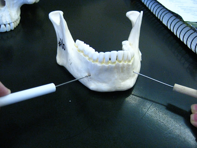
Human Skull - mental foramen (of mandible)

 
Both probes are pointing to a mental foramen (hole) on the anterior surface of the mandible.

Human Skull - coronoid process (of mandible)

The probe is pointing to the coronoid process of the mandible, which is located directly anterior to the other bony projection of the mandible (on each side) known as the mandibular condyle.

Human Skull - mandibular condyle (of mandible)

The probe is pointing to the manibular condyle, the posterior-most projection on the mandible. The mandibular condyle articulates with the cranium (i.e. skull without a mandible) at the mandibular fossa of the temporal bone.

Human Skull - ramus (of mandible)

The area of bone between the probes is the "ramus" of the mandible.

Human Skull - body (of mandible)

The metallic part of the probe is positioned along the area of the "body" of the mandible.

Human Skull - mandible

General view of the mandible (lower jaw or jaw bone).

Human Skull - lateral masses (of ethmoid bone)

Lateral/Anterior view of the skull: The probe is pointing to approximately where the lateral mass that makes up one side of the ethmoid bone is. The ethmoid bone is posterior to the nasal cavity, thus some aspects of it (like the lateral masses) are not visible in this photo.

Human Skull - nasal conchae (of ethmoid bone)

Anterior view, photo 1: The probes are pointing to the 2 superior nasal conchae (projections of bone coming off of the side walls ("lateral masses") of the nasal cavity (of ethmoid bone).
Anterior view, photo 2: The probes are pointing to the 2 inferior nasal conchae of ethmoid bone.

In total, there are 4 nasal conchae visible between these 2 photos.

Human Skull - perpendicular plate (of ethmoid bone)

The probe is pointing to the perpendicular plate (aka "vertical plate") of the ethmoid bone, visible from an anterior view of the skull through the nasal cavity. "Perpendicular" refers to the orientation of the perpendicular plate relative to the cribiform plate (aka "horizontal lamina") of the ethmoid bone.

Human Skull - cribiform plate (of ethmoid bone)

Interior top-down view: The probe is pointing to the cribiform plate (aka "horizontal lamina") that forms the horizontal "roof" of the ethmoid bone. The cribiform plate is barely visible from this view, however it is easy to identify because it is perpendicular to the vertical projection attached to it (crista galli). Memory hook: "The CRIB-iform plate is a nice place for a rooster (crista galli) to rest".

Human Skull - crista galli (of ethmoid bone)

Interior top-down view: The probe is pointing to crista galli, or "crest of the cock (rooster)", which is located perpendicular to the cribiform plate and projects upward from it.

Human Skull - ethmoid bone (calvarial bone #8)

The probe is pointing to the general area that the ethmoid bone is found in if we were to remove the superficial layer of bone to reveal the nasal cavity inside of the skull that contains the ethmoid bone. The ethmoid is the bone that separates the nasal cavity from the brain. The ethmoid is located anterior to (in front of) the sphenoid bone, and consists of 2 masses--one on each side of the nasal cavity--which are joined horizontally by 2 "cribiform plates".

Here is a picture of the ethmoid bone by itself (posterior view) from "A Brief Atlas of the Human Body" (2e).

Human Skull - foramen spinosum (of sphenoid bone)

 Interior top-down view: the probe is pointing through foramen spinosum, which is the very small circular hole immediately posterior to foramen ovale.

Underside of skull view: the probe is pointing to foramen spinosum, which is, again, directly posterior to foramen ovale.

Human Skull - foramen ovale (of sphenoid bone)

The probe is pointing through foramen ovale of the sphenoid bone (interior top-down view). Foramen ovale is a very circular-looking hole that is located anterior to a very small hole directly beside it (foramen spinosum). Memory hook: Foramen O-vale is very "O-bviously" in the middle cranial fossa ("angel wings" fossa) of the cranial floor.


Alternate photo: The probe is pointing through foramen ovale (underside view of skull), which is located lateral to foramen lacerum.

Tuesday, September 27, 2011

Human Skull - foramen rotundum (of sphenoid bone)

The probe is pointing through foramen rotundum, a circular hole in the sphenoid bone. Foramen rotundum is located directly inferior to the "main" ridge of the lesser wing (i.e. not the holes that are directly under the small, pointed projections coming off of the lesser wings).

Human Skull - optic canals (of sphenoid bone)

The probe is pointing through an optic canal located in the sphenoid bone of the skull. More specifically, the optic canals are found directly inferior to the small projections (clinoid processes) coming off of the lesser wings, or directly superior and slightly anterior to the sella turcica. The optic canal transmits the optic nerve and ophthalmic artery into the orbital cavity ("orbit") or eye socket.

Human Skull - lesser wings (of sphenoid bone)

The probe is pointing to a lesser wing of the sphenoid bone from an interior top-down view of the skull. The lesser wings are located superior to their respective greater wing. In terms of the "bat" analogy for the sphenoid bone (when viewed from an anterior or posterior view), the lesser wings would appear to be the anatomical "ears" of the bat/sphenoid bone.

Human Skull - sella turcica (of sphenoid bone)

The probe is pointing to the sella turcica of the sphenoid bone. The sella turcica or "Turkish chair" is a horse saddle-shaped dip in the sphenoid bone that also includes the dorsum sellae, which is the bony projection/ridge that the probe is specifically pointing to.

Human Skull - inferior orbital fissure (of sphenoid bone)

The probe is pointing through an inferior orbital fissure at the posterior base of one of the eye sockets. Each eye socket has an inferior orbital fissure with a corresponding superior orbital fissure located directly above the inferior orbital fissure.

Human Skull - superior orbital fissures (of sphenoid bone)

The probe is pointing through a superior orbital fissure of one of the eye sockets. There is one superior orbital fissure per eye socket, and it is located above the inferior orbital fissure at the posterior base of the eye socket.

Human Skull - pterygoid process (of sphenoid bone)

The probe is pointing to the pterygoid process of the sphenoid bone from a "side" or lateral view of the skull. Each of the two pterygoid processes resembles an "ankle" of the sphenoid bone (aka "bat") when the sphenoid bone is viewed from  a posterior or anterior view

Human Skull - greater wings (of sphenoid bone)

The probe is pointing to a greater wing of the sphenoid bone from an interior top-down view of the skull. In terms of the "bat" analogy used for the appearance of the sphenoid bone, the greater wings resemble the anatomical "main wings" of the bat/sphenoid bone from an anterior or posterior view. The whole sphenoid bone is not visible here--for example, the pterygoid process is not visible from this angle.

Human Skull - sphenoid bone (calvarial bone # 7)

The probe is pointing to the general area where the sphenoid bone (aka "bat") is found in the skull (it is inside the skull, most of it is not visible from this anterior view).

Human Skull - superior nuchal line (of occipital bone)

The probe is pointing along the superior nuchal line, which is located directly superior to the visible ridge (known as the "external occipital protuberance", which is a useful landmark) below it.

Bone terms:
line - is slight, long, linear bump or ridge; it is like a "crest" but does not protrude out as far.


Human Skull - condyloid canal (of occipital bone)

The probes are pointing to the condyloid canals on the underside of the skull, located contralateral to the foramen magnum and posterior to the occipital condyles.

Human Skull - hypoglossal canal (of occipital bone)

The probe is pointing through the hypoglossal canal, which is located directly superior and slightly anterior to its corresponding occipital condyle on each side of the skull. The hypoglossal canals are, in general, located contralateral to the foramen magnum, and serve to transmit the nerve that supplies the muscles ("motor innervation") of the tongue.

Human Skull - occipital condyle (of occipital bone)

The probe is pointing to an occipital condyle on the underside of the skull (base). The two occipital condyles are large rounded kidney-shaped projections of occipital bone that are located contralateral to the foramen magnum and that articulate with the superior facets of the "Atlas" (C1) cervical vertebra.

Bone Terms:
condyle - a smooth, articulating surface (a joint) that slightly curved, typically. It is often covered with articular cartilage that is smooth and "slippery" to reduce friction between bone surfaces during movement.

Human Skull - foramen magnum (of occipital bone)

The probe is pointing to the foramen magnum, or "great hole" of the underside of the skull. Foramen magnum is the hole through which the medulla oblongata (an extension of the spinal cord) enters and exits the skull.

Human Skull - lambdoid suture (of occipital bone)

The probes are pointing to the lambdoid suture, which is named for the "lambda" shape it forms with the sagittal suture (i.e. the vertical suture in this photo).


Human Skull - occipital bone (calvarial bone # 6)

The probe is pointing to the occiptal bone (in general).

Human Skull - petrous part (of temporal bone)

The probe is pointing to the petrous part, which houses the internal acoustic meatus. The petrous part visible in this interior top-down view is the large ridge of bone that separates the posterior cranial fossa from the middle cranial fossa.

Human Skull - foramen lacerum (of temporal bone)

The probe is pointing to foramen lacerum, which is a "lacerated"/triangular-shaped "shredded" hole on each side of the base of the skull. That is, the foramina lacerum are located contralateral to the center portion of the base of the skull known as "pharyngeal tubercle" of the occipital bone.

Human Skull - internal acoustic meatus (of temporal bone)

The probe is pointing to the internal acoustic meatus, which is also known as the internal auditory canal. The whole internal acoustic meatus is located inside the petrous part of a temporal bone, however, note that the opening is located near the center of the posterior surface of the petrous part, superior to the jugular foramen (interior top-down view).

Human Skull - carotid canal (of temporal bone)

The probe is pointing to/through the carotid canal on the underside of the skull. The carotid canal has a circular, smooth opening and is located immediately anterior to the jugular foramen.

Alternate view: here is an interior top-down view of the jugular foramen not taken from class. From this view, it is clear that the carotid canal is located anterior to the petrous part of temporal bone.

Monday, September 26, 2011

Human Skull - jugular foramen (of temporal bone)

The probe is pointing to a jugular foramen, a relatively large, weirdly shaped (i.e. non-circular) opening at the base of the skull (view is from the underside of the skull). There is one jugular foramen on each side of the base of the skull, contralateral to foramen magnum. Jugular foramen can be distinguished from other foramina/holes by remembering that the jugular foramina are located immediately lateral to the occipital condyles (the 2 obvious "nubs" on the foramen magnum).

Alternate view: here is an interior top-down view of the jugular foramen not taken from class. From this view, it is clear that the jugular foramen is located posterior to the petrous part of temporal bone.


Human Skull - mastoid process (of temporal bone)

The probe is pointing to the mastoid process, which looks like a rounded cone protruding from the underside of the skull. The mastoid process is located almost directly behind (posterior to) the external auditory meatus, and is also lateral to the styloid process (left*) of a temporal bone.

*see previous post on "styloid process" for a clearer visual example of the styloid and mastoid processes side-by-side.

Human Skull - styloid proces (of temporal bone)

The probe is pointing to the styloid process, which is a very pointy, stylus-shaped piece of bone. The styloid process in this photo is view from the underside of the skull, and it can be easily seen as the pointed protuberance directly below (inferior to) the external auditory meatus of a temporal bone.

FYI - the mastoid process is located directly lateral to the styloid process. In the photo above, the mastoid process is the "bump" immediately to the right of the styloid process (and the probe). This is a fairly clear picture of the two processes side-by-side.


Human Skull - external auditory meatus (of temporal bone)

The probe is pointing to the external auditory meatus ("ear canal"), which is a (mostly) bony tube that runs from the outer ear to the middle ear.

Bone terms:
meatus - can refer to: (1) the opening to a canal or tube, or (2) the canal or tube itself.


Human Skull - mandibular fossa (of temporal bone)

The probe is pointing to the mandibular fossa, which is the shallow"dip" in a temporal bone that articulates with the mandibular condyle (i.e. where the lower jaw fits into the skull to form a joint).

Bone terms:
fossa - a shallow, basin-like depression in a bone; a concave (cup-like) indentation or dip in a bone.

Human Skull - zygomatic process (of temporal bone)

The probe is pointing to the zygomatic process of *temporal* bone (NOT "of zygomatic bone").

Bone terms:
process - any protuberance, "bump", or "hill" of a bone; any other structure of a bone that sticks out.
a few sub-types of "process": trochanter, crest, tubercle, condyle, epicondyle, tuberosity, line, spine.
Human Skull - squamous suture (of temporal bones)

The probe is pointing to the squamous suture, which forms the top "arch" of a temporal bone, separating it from the lower border of a parietal bone.

Human Skull - temporal bones (calvarial bones # 4 and # 5)


Both pictures: the probe is pointing to a temporal bone. The skull has 2 temporal bones total, one on each side (literally) of the skull.

Human Skull - coronal suture (of parietal bones)

The probe is pointing to the coronal suture, which is another dense, fibrous joint and synarthrosis, just like the sagittal suture (previous post). The coronal suture separates the frontal bone from the 2 parietal bones. The coronal suture gets its name from being located on the "crown" (top) of the head.

Human Skull - sagittal suture (of parietal bones)

The probe is pointing to the sagittal suture, which is a dense, fibrous joint (and is a synarthrosis, because there is no mobility) between the 2 parietal bones of the skull.

Human Skull - parietal bones (calvarial bones # 2 and # 3)

Both pictures: the probe is pointing to a parietal bone. The skull has 2 parietal bones total, one on each side on the top of the skull (i.e. contralateral to the sagittal suture).